Makati

Philippines

Phone Number

0962 950 7814
(02) 8637-2360

Send Your Mail

slcelitemedicalcare@gmail.com

Hospital Equipment

Empowering Care with Precision, Safety, and Innovation

The hospital has a complete set of modern diagnosis and treatment equipment such as positron emission Tomography System (PET-CT), Radiotherapy System Linear Accelerator (LA), 3.0T magnetic resonance imaging system (MRI), single-photon emission computed tomography system (SPECT/CT), vascular interventional therapy system (digital subtraction/DSA), 3D thoracic laparoscopic system, digital operating room and other modern diagnosis and treatment equipment.

Positron Emission Tomography - Computed Tomography

A PET-CT (Positron Emission Tomography – Computed Tomography) system is an advanced medical imaging technology that combines two scanning techniques to provide detailed information about both structure (anatomy) and function (metabolism) in a single scan.

How It Works:
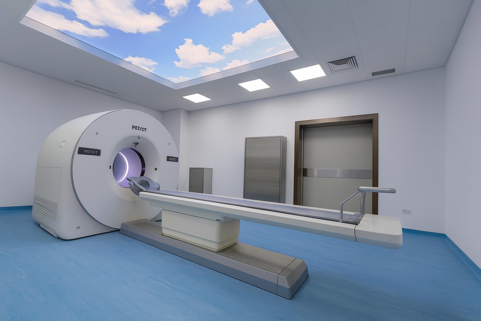
  • PET Scan:
    • Detects metabolic activity using a radioactive tracer (e.g., FDG, a glucose analog).
    • Cancer cells, which consume more glucose, “light up” on the scan.
  • CT scan:
    • Provides 3D X-ray images of bones, organs, and tissues for precise anatomical localization.
  • Fusion Imaging:
    • Combines PET (function) + CT (structure) to pinpoint abnormalities (e.g., tumors, inflammation) with millimeter accuracy.

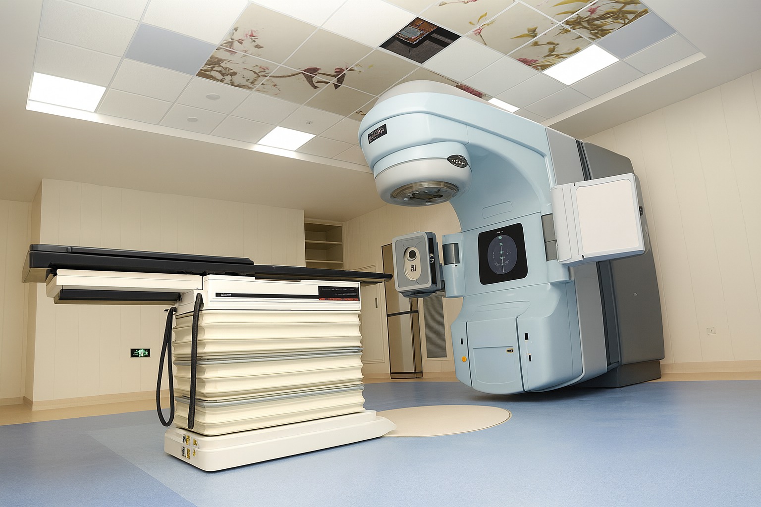
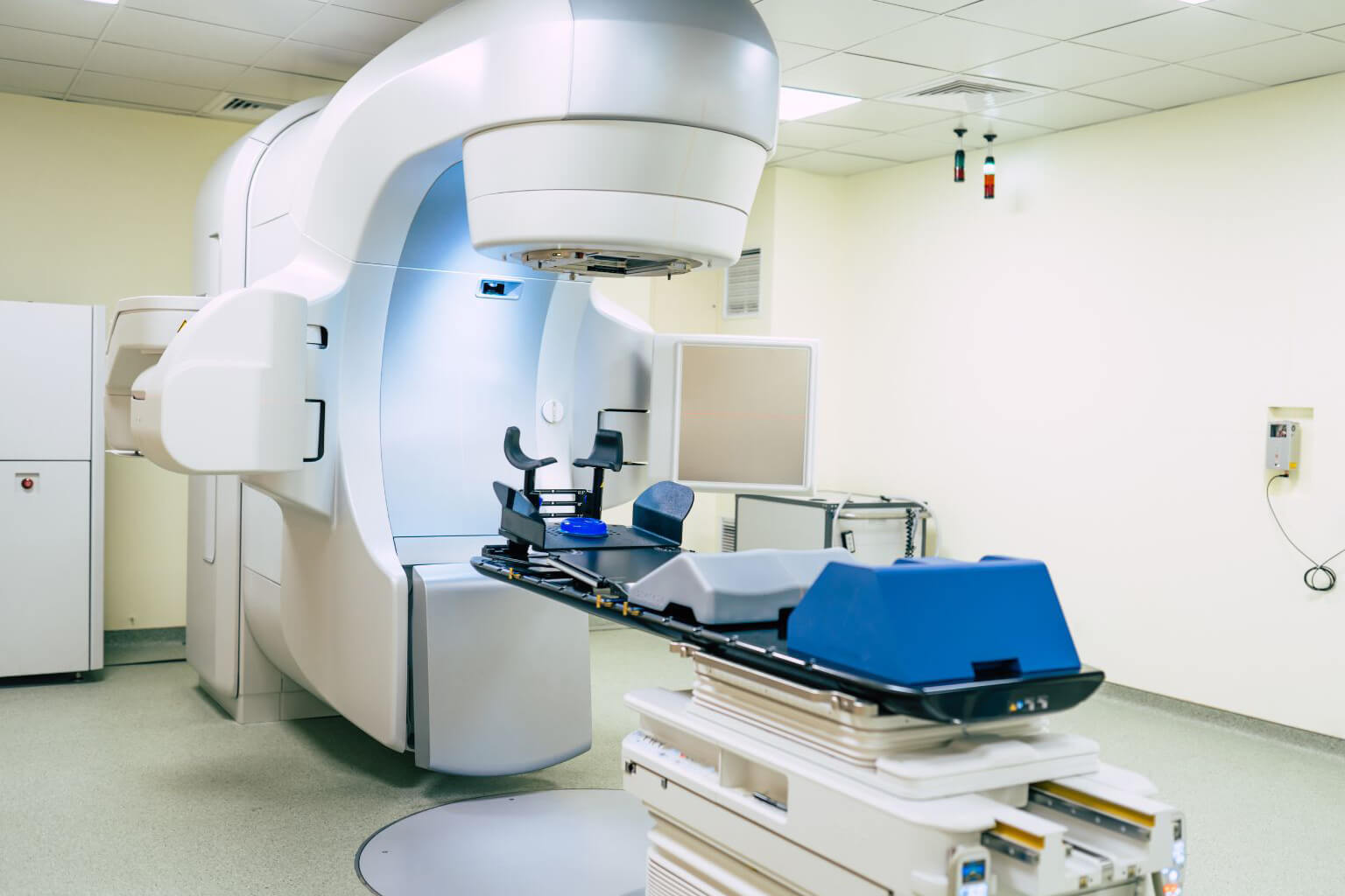
Linear Accelerator

A Linear Accelerator (LINAC) is the most advanced and widely used radiotherapy system for treating cancer. It delivers high-energy X-rays or electrons precisely to tumors while minimizing damage to surrounding healthy tissues.

How a LINAC Works:

  • Beam Generation:
    • Electrons are accelerated to near light-speed in a linear tube using microwave technology.
    • These electrons either:
      • Strike a metal target to produce high-energy X-rays (for deep tumors).
      • Or are used directly as electron beams (for shallow cancers like skin tumors).
  • Precision Targeting:
    • Uses 3D imaging (CT/MRI-guided) to map the tumor’s exact size/shape.
    • Shaped by a multi-leaf collimator (MLC) to match the tumor’s contours (within 1mm accuracy).
  • Treatment Delivery:
    • The patient lies on a movable couch while the LINAC’s gantry rotates 360° to attack tumors from multiple angles.
    • Intensity-Modulated Radiotherapy (IMRT) or Stereotactic Radiosurgery (SRS) adjusts beam strength in real time.

Tesla Magnetic Resonance Imaging

A 3.0 Tesla Magnetic Resonance Imaging (MRI) system is a high-field, advanced medical imaging device that uses powerful magnetic fields (twice as strong as standard 1.5T MRI) and radio waves to produce exceptionally detailed, high-resolution images of the body’s internal structures without radiation.

Key Features of 3.0T MRI:
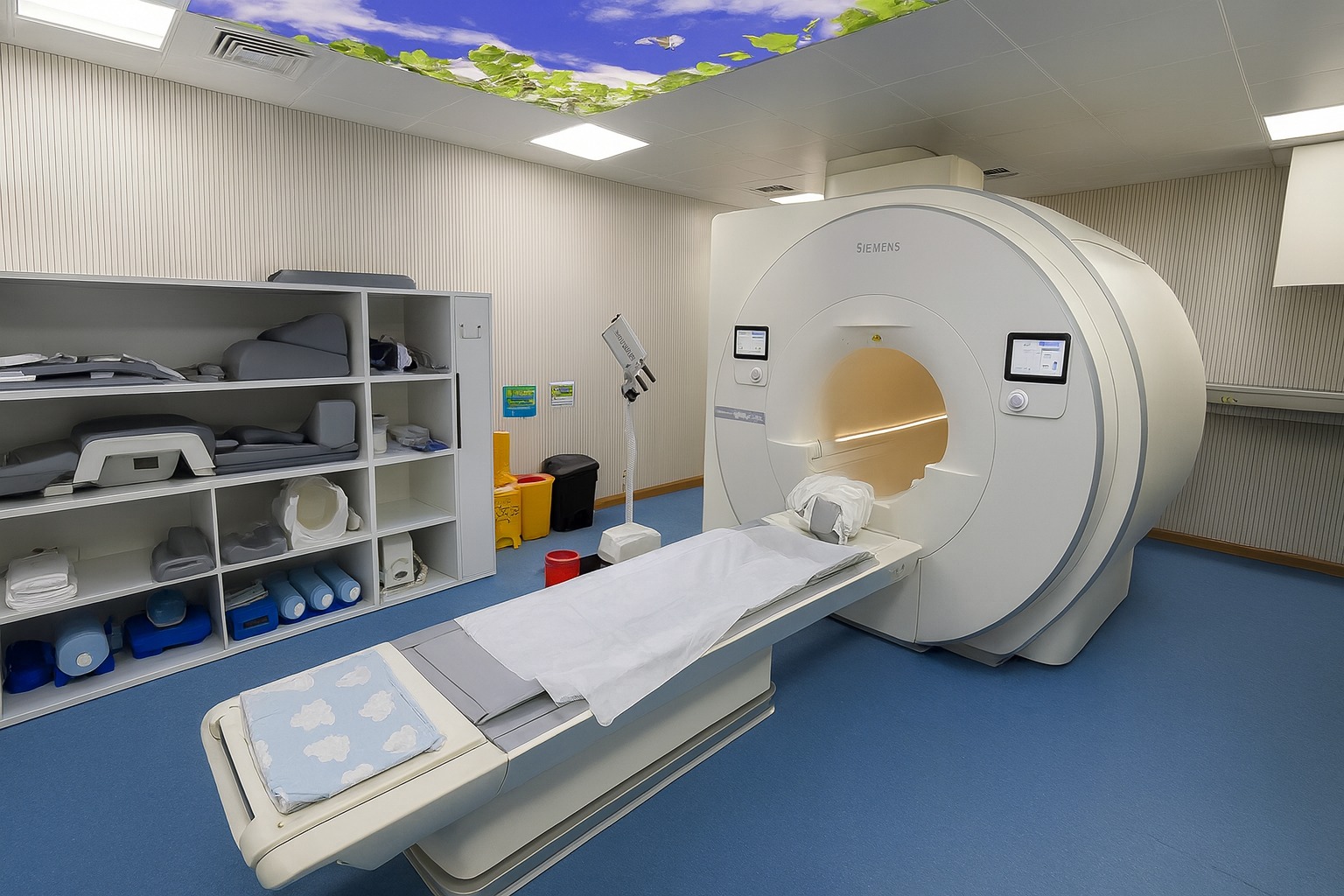
  • Ultra-High Magnetic Field (3 Tesla)
    • 2x stronger than conventional 1.5T MRI → Sharper images, especially for small structures (e.g., nerves, blood vessels, brain lesions).
    • Faster scan times (reduces motion artifacts).
  • Superior Image Quality
    • Higher signal-to-noise ratio (SNR) → Clearer soft-tissue contrast for:
      • Brain (stroke, tumors, multiple sclerosis)
      • Musculoskeletal (torn ligaments, cartilage)
      • Cardiac (heart muscle viability)
      • Cancer staging (prostate, breast, liver)
  • Advanced Applications
    • Functional MRI (fMRI): Maps brain activity in real time.
    • Diffusion Tensor Imaging (DTI): Tracks nerve fiber pathways.
      Magnetic Resonance
    • Angiography (MRA): Visualizes blood vessels without contrast dye.
    • Spectroscopy: Detects biochemical changes in tissues (e.g., tumor metabolism).

How It Works:

  1. The patient lies inside a cylindrical magnet, aligning hydrogen atoms in the body.
  2. Radiofrequency pulses disrupt this alignment; as atoms return to normal, they emit signals.
  3. A computer converts these signals into 3D cross-sectional images (slices as thin as 1mm).

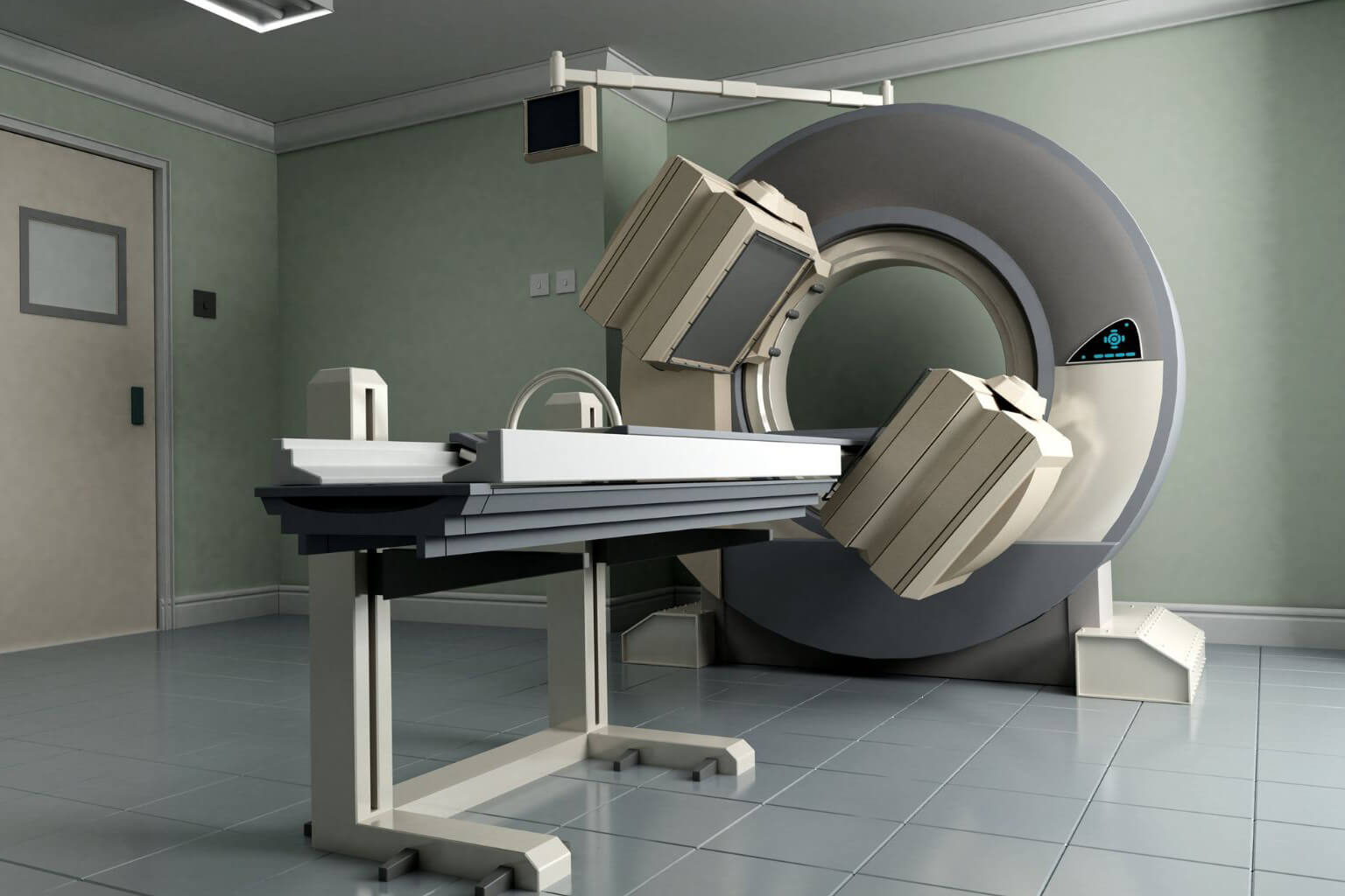
Single-Photon Emission Computed Tomography/Computed Tomography

A Single-Photon Emission Computed Tomography/Computed Tomography (SPECT/CT) system is a hybrid medical imaging device that combines functional (SPECT) and anatomical (CT) imaging to provide detailed 3D maps of physiological processes in the body. It is widely used in oncology, cardiology, neurology, and orthopedics.

How SPECT/CT Works:

  • SPECT (Functional Imaging):
    • A radioactive tracer (e.g., Technetium-99m, Iodine-123) is injected into the patient.
    • The tracer emits gamma rays as it accumulates in target tissues (e.g., tumors, bones, heart muscle).
    • A gamma camera rotates around the patient, detecting these rays to create 3D metabolic images.
  • CT (Anatomical Imaging):
    • X-rays generate high-resolution structural images of bones, organs, and blood vessels.
  • Image Fusion:
    • Combines SPECT (metabolic activity) + CT (anatomy) to precisely localize abnormalities (e.g., a tumor’s exact size/location).

Vascular Interventional Therapy System

A Vascular Interventional Therapy System (Digital Subtraction Angiography, DSA) is an advanced medical imaging technology used to diagnose and treat vascular (blood vessel) diseases in real time. It combines high-resolution X-ray imaging with catheter-based minimally invasive procedures to visualize and repair blood vessels with pinpoint accuracy.

How DSA Works:
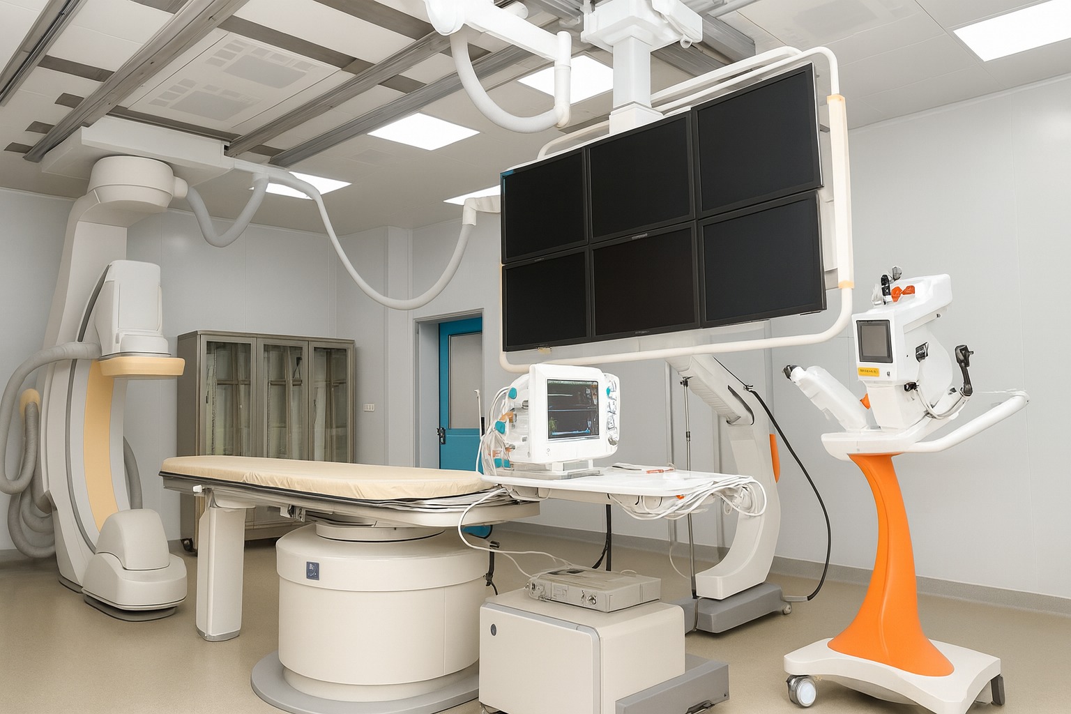
  • Image Acquisition:
    • Baseline X-ray: First, a standard X-ray image is taken.
    • Contrast Injection: A contrast dye is injected into the blood vessels via a catheter.
    • Digital Subtraction: The system removes (“subtracts”) bone and soft tissue from the images, leaving only a clear view of the blood vessels (angiogram).
  • Real-Time Guidance:
    • The live X-ray feed helps doctors navigate catheters/wires to:
    • Diagnose blockages, aneurysms, or malformations.
    • Treat by deploying stents, coils, or dissolving clots.
  • Therapeutic Procedures:
    • Angioplasty: Opens narrowed arteries with a balloon.
    • Embolization: Blocks abnormal vessels (e.g., tumors, aneurysms).
    • Thrombectomy: Removes life-threatening blood clots.

Thoracic Laparoscopic System

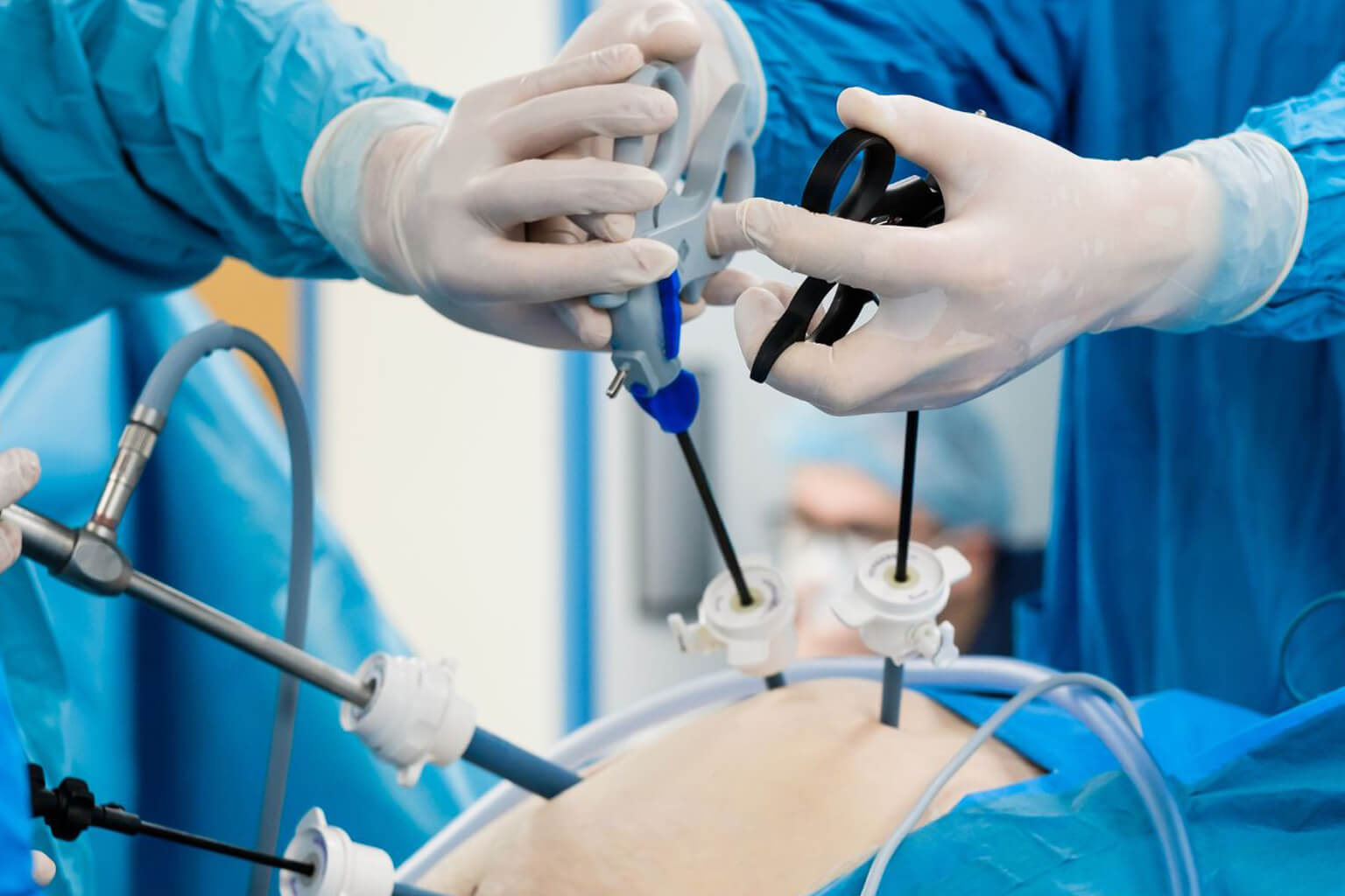
A 3D Thoracic Laparoscopic System is an advanced minimally invasive surgical platform that combines high-definition 3D visualization with specialized instruments to perform precise surgeries in the chest (thoracic cavity) through tiny incisions. It is commonly used for lung, esophageal, and mediastinal procedures.

Key Components & How It Works:

  • 3D High-Resolution Camera:
    • Dual-lens endoscope captures stereoscopic images, giving surgeons depth perception (like human binocular vision).
    • 4K/8K resolution enhances detail of delicate tissues (e.g., blood vessels, nerves).
  • Thoracic Laparoscopic Instruments:
    • Thin, articulated tools (e.g., graspers, staplers) inserted through 5–10mm ports.
    • Wristed movements mimic human hand dexterity in tight spaces.
  • Integrated Imaging & Navigation:
    • Real-time fusion with preoperative CT/MRI for tumor localization.
    • Fluorescence imaging (e.g., ICG dye) highlights critical structures.